
- පුවත්
- |
- පර්යේෂණ
New Study unpacks Cerebral Cortex Morphometry and Relaxometry in Male Children With Fragile X Syndrome and Autism
Published: 31 මැයි 2026
For the complete article by Guerrero-Gonzalez and others, please click here.
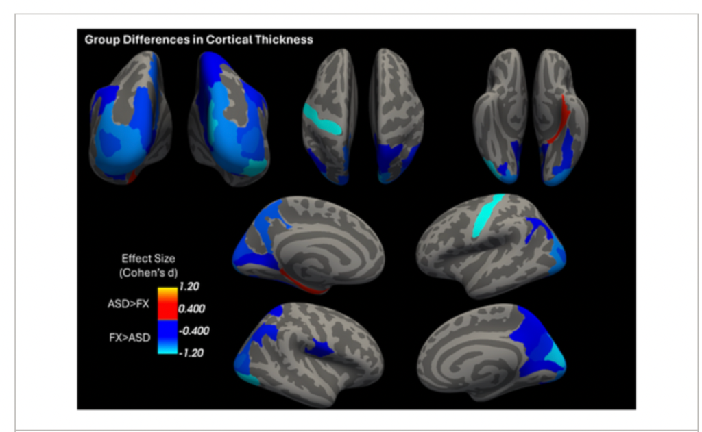
Pictured above, Significant group differences in cortical thickness between FXS and ASD (all credit to the authors of the original article). These were found primarily within occipital, parietal, and temporal regions
Cortical thickness is a macro-structural measure and therefore includes contributions of multiple microstructural features including myelination and cell density. Previous studies have proposed that cortical thickness may have an association with age and cognitive functioning. It is difficult to spot differences in the way cortical thickness impacts males living with FXS and those who live with other intellectual conditions. Employing a motion-robust quantitative brain imaging method to enable accurate measuring of cortical thickness, the authors conducted a study comparing the effects of cortical thickness in 50 male participants with Autism Spectrum Disorder (ASD) and 11 with FXS.
About 30-50% of males living with FXS meet the criteria for ASD, making FXS the most studied mono-genetic model of ASD. Previous studies suggest that there are clear differences between the two conditions. For example:
- FXS combines rather preserved receptive vocabulary with greater gaze avoidance and social anxiety in social communication.
- Individuals living with FXS omit more function words and show greater narrative coherence than those with ASD.
- Individuals with FXS display high-frequency motor stereotypies but fewer insistence-on-sameness rituals.
- FXS displays more pervasive auditory, tactile and visual hypersensitivity.
- FXS shows more severe attention-inhibition deficiencies compared to ASD.
- Smaller temporal limbic volumes and larger caudate nuclei are observed in FXS.
The study contributes to growing studies on cortical thickness by engaging in an in-depth analysis of findings pertaining to FXS.
- Individuals with FXS showed statistically increased cortical thickness across multiple visual-processing regions suggesting alterations in visual cortex development that may underlie the sensory processing features. The increased cortical thickness in visual regions may reflect impaired synaptic pruning mechanisms, as FMRP normally regulates the translation of proteins essential for synaptic refinement and elimination of inappropriate connections.
- Critical components of the default mode network (DMN) which are involved in visuospatial attention, spatial working memory, and self-referential processing appear to be altered due to cortical thickness. The precuneus, in particular, serves as a hub for integrating information across brain networks and is essential for consciousness and self-awareness.The increased cortical thickness in these regions in FXS may contribute to the altered spatial processing and attention difficulties observed in this condition.
- There was a positive relationship between age and cortical thickness in the parahippocampal gyrus in both groups, contrasting with the inverse relationships observed elsewhere. This suggests active developmental processes continuing through adolescence.
- The significant age-by-group interaction in the central sulcus, with a stronger inverse relationship in the FXS group, suggests altered cross-sectional age-related associations in sensorimotor regions. The central sulcus contains the primary motor and somatosensory cortices, and the stronger age-related thinning in FXS may reflect accelerated or dysregulated pruning processes in these regions. This finding is consistent with the motor coordination difficulties and sensory processing differences commonly observed in FXS.
The authors highlight that understanding differences in developmental trajectories and brain structural differences between FXS and autism may have potentially important implications for intervention strategies. If more studies incorporating wider age ranges and gender emerge, such patterns of group differences may help identify developmental windows during which imaging markers are most informative for linking brain features to symptom profiles and treatment response.

